
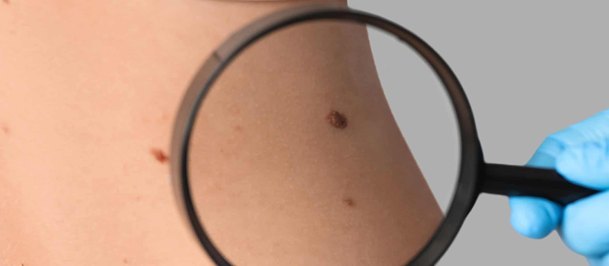
Moles, which are often pink or brown, oval or round, can appear later as well as congenital. However, the cause of the molecular change that plays a role in the formation of moles that can be seen in every part of the body is not fully known.
Most of the moles are benign, while some of them may develop skin cancer called ‘melanoma’. Those with light skin and eye color, red-blond hair, easily burning or difficult tanning, prone to freckling, prolonged or intermittent intense sun exposure, abnormal (atypical) looking (dysplastic) nevus, those with a large number of congenital moles and those with a family history of melonoma have a greater risk for skin cancer.
Color, shape and size changes in moles should be followed. If the edges of the mole are large and irregular, the color changes, there are growths, symptoms such as burning, itching, pain, bleeding occur in the mole, a dermatologist should be consulted as soon as possible. It is also important that people with many and large moles and people with melonoma in their family see a specialist again. Ultraviolet rays can cause an increase in the number of moles and cancer. For this reason, sunscreens with a high protection factor should be used in summer and winter.
‘Melanoma’, whose risk of being caught increases every year, is a type of skin cancer that has the potential to spread fast enough to be life-threatening and mole examination is very important for early diagnosis of the disease. One of the biggest misconceptions about moles in society; is that when the mole is removed, it will turn into cancer. However, removing the mole does not cause it to turn into cancer, on the contrary, it is appropriate to remove the moles that your doctor finds risky.
Moles can also be removed when they are not wanted for aesthetic reasons. The removal alone should be done by a plastic surgeon.
As well as excessive exposure of moles on the body to sunlight, entering the solarium, excessive and incorrect use of bad cosmetic products also leads to differentiation in moles. Conditions such as mole plucking and cutting are definitely not recommended as they can cause different structural changes in the mole. Meat moles, which are a kind of skin attachment, do not have any danger for the person and are not likely to turn into cancer. Meat moles, which are more common in areas such as neck and armpits, may increase with age, weight and pregnancy. If they are aesthetically disturbing to the person or traumatized by being attached to clothes and accessories used, they should be removed.
During the examination of moles, an instrument called digital dermotoscopy is used. This device enlarges the moles 20-40 times and takes a dermoscopic image of the mole and records it on the computer. Thus, by comparing the image obtained at the next control of the person, suspicious changes in moles can be detected.
Digital dermatoscopy mathematically calculates the suspicious changes observed in the mole and creates a score indicating the risk of melonoma disease. Thus, the follow-up and treatment of the mole can be done more healthily. While the chance of diagnosing early stage melanoma with the naked eye is 60 percent, it increases to 90 percent with digital dermatoscopic examination. While 30 percent of the ‘Malignant Melanoma’ lesions, which are one of the fastest increasing cancer types in the world, develop on a mole, 70 percent occur on a normal skin background.
‘Malignant Melanoma’, which can be effectively treated when diagnosed at an early stage, eliminates the chance of treatment by rapidly spreading to regional lymph nodes and then to the whole body if it cannot be diagnosed in this period.
Each individual is advised to examine their moles at monthly intervals. Asymmetry, boundary irregularity, discoloration (the mole contains more than one color), significant growth, inflammatory reaction or bleeding in the mole detected during this examination may indicate skin cancer. The patient can be seen with his doctor by comparing these changes with the old findings at the same time on the screen. People should have mole follow-up as soon as they suspect moles and increase the chance of early diagnosis against melanoma risk.
Mole removal surgery is performed under local anesthesia. In this way, the patient does not feel any pain or suffering during surgery. Anesthesia is applied around and under the mole with small needles. After the mass removal operation, pathological examination of the mole should also be performed. The procedure takes 5-10 minutes. The wound formed after this operation is closed by suturing. After a good operation, the scar remains very little. In general, there are a few millimeters more scars than the size of the mole.
Over time, it also becomes imperceptible with the maturation of the scar. After the procedures, the wound is dressed and closed. It does not need to be supervised in any way after surgery. The patient can immediately return to social and work life. Mole removal is a fairly safe procedure. You may experience a slight redness or itching sensation in the area where the mole was removed, which will disappear within a few days. The risk of infection is quite low. The effect of local anesthesia lasts for several hours. After the effect has passed, complaints in the form of mild burning-pain can be controlled with simple painkillers. After the first two days, the wound can be washed. Your plastic surgeon will give you various recommendations to leave less. It is also important to stay away from the sun’s rays.
- Plastic Surgery
- Mommy Makeover
- Non-Surgical Facial Rejuvenation
- Leg Lift Surgery
- Bichectomy
- Lower Face Rejuvenation
- Rhinoplasty
- Jaw Tip Aesthetics
- Genital Aesthetics
- Blepharoplasty (Eyelid Surgery)
- Inward Nipple Operation
- Gynecomastia
- Hip Butt Aesthetics
- Abdominoplasty (Tummy Lift)
- Eye Brow Lift
- Otoplasty (Ear Aesthetics)
- Arm Stretching
- Liposuction
- Breast Augmentation Operation
- Breast Lifting Operation
- Reduction Mammaplasty
- Operations After Bariatric Surgery
- Mid Face Lift
- Fat Injections to the Face
- Non-Surgical Aesthetics
- Plastic surgery