
The skin, which is the largest organ in the body, has many tasks. The skin protects the body against possible injuries, prevents fluid loss and helps the body provide vitamin D. In addition, it protects against both microorganisms and harmful ultraviolet (UV) rays and helps control body temperature.
Skin cancer is a malignant growth that occurs on the skin for many reasons. Skin cancers can be grouped into two main groups as melanoma and non-melanoma skin cancers.
Melonoma Skin Cancer
Melanoma is a skin cancer that starts in cells called melanocytes that give the skin its color. It is called malignant melanoma or cutaneous (skin-borne) melanoma. Melanoma, the most malignant of the skin cancers, accounts for about 4 percent of all skin cancers. In melanoma, the tumor is usually brown or black in color, as most of its cells continue to produce melanin. However, some melanomas do not produce melanin. In this case, the tumor can be seen as pink, yellowish-brown or even white.
Melanoma can develop on moles that are present at birth or appear later in the skin. The scalp can be seen anywhere in the skin covering the body, including the soles of the feet. In the neck and back areas in men; It is common in women on the legs, neck and face. But cancer can also develop on the palms, soles of the feet, nail bed. In addition to all these, although rare, it can also occur in the eye, mouth, genital or anal area.
While it is very rare for non-melanoma cancers to spread to other parts of the body, melanomas often spread to surrounding tissue and other parts of the body. Since melanoma, which is a treatable type of cancer when diagnosed at an early stage, spreads rapidly, the chance of treatment in the late period is low. Other (Non-Melanoma) Skin Cancers
Basal cell and squamous cell carcinomas are the most common tumors of this group. Mercel cell carcinoma and sebaceous carcinoma, which are rarer, are also included in this classification. It is mostly seen in areas that are overexposed to the sun, such as the head, neck and arms. However, this is not a rule.
Basal Cell Carcinoma: It is the most common skin cancer. It accounts for about 80 percent of all skin cancers. They are traced as small, slow-moving, bright, pink or red piles. If you leave it alone, they tend to crust, bleed and turn into ulcers. Basal cell cancers that originate from cells in the epidermis, the outermost layer of the skin, grow slowly and rarely spread. It occurs more in people with light skin and colored eyes. Squamous Cell Carcinoma: It is the second most common skin cancer. These cancers, which originate from squamous cells in the epidermis, are responsible for 16 percent of all skin cancers. It may look like a growing tuber. It often has a hard, flaky or crusty surface. It may also have a flat, red-spotted appearance that develops slowly.
Aknitic keratosis is also known as solar keratosis. It is usually considered a precancerous condition and occurs as a result of excessive exposure to the sun. Aknitic keratosis is small, hard or flaky spots, usually pink-red or skin colored. It usually starts on the face, ears, above the hands and arms. However, it can also occur in other parts of the body exposed to the sun. Aknitic keratosis is usually numerous. Some may develop within squamous cell cancers. Others remain the same or go it alone. Factors That Cause Skin Cancer
The most important are UVA and UVB rays. With prolonged exposure to ultraviolet rays, the incidence of all skin cancers, including melanoma, is increased. Likewise, people who often go to the solarium to tan have a higher risk of melanoma skin cancer. Lamps used for tanning in solariums should have labels that read “Constant exposure to UV rays can cause premature aging of the skin and skin cancer.” In addition, it is thought that putting another label that says “regular doctor control is necessary for skin cancer” for users who are constantly exposed to these rays may be of a warning nature.
Moles in our body are benign tumors and are formed not only at birth but also during childhood and adolescence. Most moles never cause problems. However, people with a large number of moles have a greater risk of developing melanoma. Dysplastic Nevuses often look like normal moles but carry some features of melanoma. They are usually larger than other moles and their shape or color is unusual and most do not turn into cancer.
It has been determined that the risk of developing melanoma in such moles that exist with birth called congenital (Congenital) Melanocytic Nevus is between 0-10% depending on the size of the nevus. People with a large congenital melanocytic nevus have a greater risk of developing melanoma.
People with white skin and light hair have a 10 times higher risk of melanoma than people with black skin. People with red and blond hair, white skin, blue or green eyes, or freckles have an increased risk of skin cancer.
Dealing with welding and metalwork has been shown to increase the risk of melanoma in the eyes. UV rays exposed during phototherapy (light therapy) applied to people with some skin diseases such as psoriasis increase the risk of squamous cell skin cancer.
People who have melanoma in one or more first-degree relatives (mothers, siblings or children) have a higher risk of skin cancer. Approximately all people with melanoma have the same disease in their family. One reason for the increased risk may be frequent sun exposure as a result of similarity in lifestyle within the family, the presence of sensitive light-skinned family members in the majority, or both of these factors. In addition, gene mutation as a result of changes in genes in the family may be a factor in melanoma risk. Gene mutations are found in approximately -40 of people with a high rate of melanoma in their family.
Xeroderma pigmentosum (XP) is an inherited and rare disease that normally develops as a result of damage to enzymes responsible for repairing DNA. People with XP may develop many skin cancers, including melanoma, in sun-exposed areas of their skin because they have less ability to repair DNA damage caused by the sun.
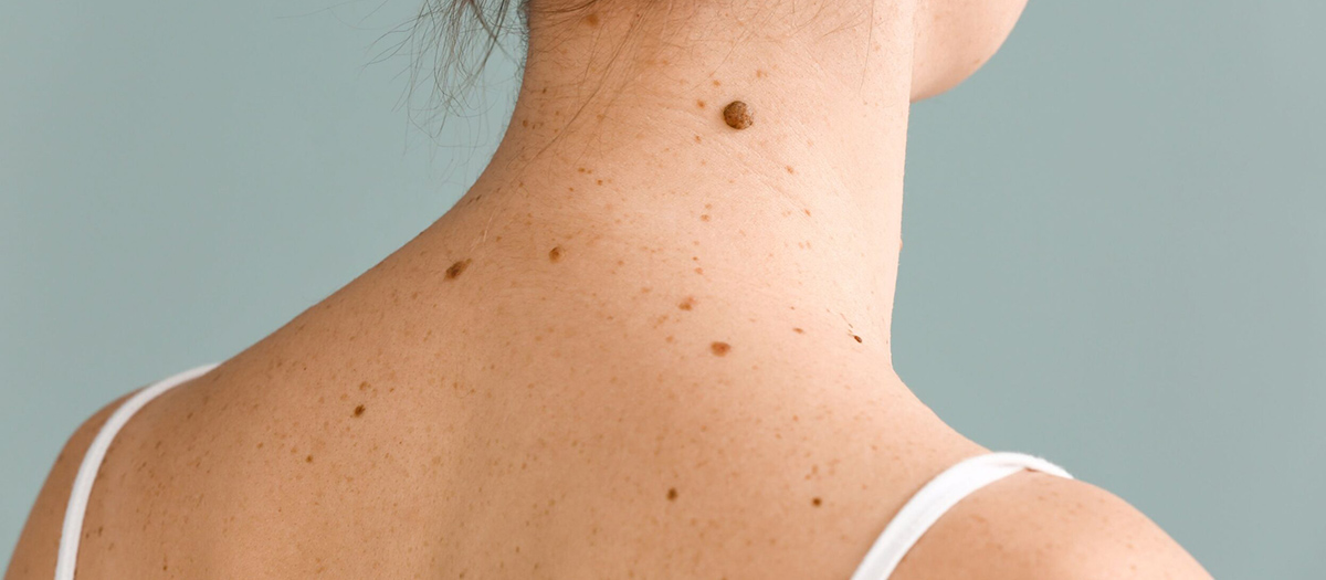
Normal moles are usually brown, tan or black spots on the skin. It can be flat or high and fluffy, round or oval and usually smaller than 6 mm. Moles can form at birth or during childhood and adolescence. New moles that appear on the body during adulthood should be checked by a doctor for a possible skin cancer. When a mole develops in the body, it will remain the same size, shape, and color for many years. Some moles will disappear spontaneously over time. Many people have moles, and most of these moles are harmless. However, when there is a change in the shape, size or color of the mole, it is important to notice this against the possible development of skin cancer.
Symptoms of Skin (Skin) Cancer
The most important symptom of skin cancer is a new spot or a change in the size, shape or color of a new spot or blemish on the skin. Other warning signs are non-healing wounds, pigments spreading beyond the stain to the surrounding skin, redness or a new swelling beyond its border, itching, tenderness or pain, change of the surface of the mole – bumping, bleeding or the appearance of a nodule or lump. It can sometimes be difficult to distinguish between a normal mole and melanoma. When such symptoms are encountered, consulting a specialist doctor without losing time will ensure that a quick solution to the health problem detected early is found.
It is very important that once a month the skin is checked by the person himself. Knowing the characteristics of moles, spots and freckles, it is important to recognize the body enough to notice a new mole or stain. Self-skin examination should be carried out in a well-lit room in front of a full-length mirror, where the whole body can be observed. By using a hand mirror, it is easier to view the blind spots of the body such as back and hips in the full-length mirror. In men, 1 in 3 melanomas is seen in the back. All areas of the body such as the back, palms, soles of the feet, scalp, eyes, nails should be checked.
A physical examination for skin cancer should first be performed by a qualified dermatologist. Using a technique called dermatoscopy, spots or moles on the skin are closely examined. Some skin cancers spread to the lymph nodes. When this happens, the affected lymph nodes may enlarge and become harder than normal. It is important that people with dysplastic nevus syndrome, who have a family history of melanoma, have regular skin examinations. Dermatoscopy is skin surface microscopy. It is used in the diagnosis of moles and other pigmented lesions. In this method, the skin surface is examined with a dermatoscope that provides a luminous magnification.
Until a few years ago, mole follow-up was provided by taking photographs of dermatoscopic images. Then, computer technology was added to this method and digital dermatoscopy was developed. With this method, the map of the moles in the body is created and their point localization is determined. Then, a dermatoscopic image is taken for each mole and recorded. This provides a chance to compare with the image to be obtained at the next check. The digital dermatoscope also creates an index showing the risk of malignant melanoma by mathematically calculating the suspicious changes observed in the mole. This index is helpful in diagnosis and treatment planning. While the chance of diagnosing early stage melanoma with the naked eye is ‘, with digital dermatoscopic examination?’ s.
If it is thought that the suspicious area may have skin cancer, a piece sample is taken from this area and examined in the laboratory, which is called a skin biopsy. A skin biopsy can be done in many different ways. The biopsy form will be decided according to the location of the suspected area in the body, its size and the suspected type of skin cancer. Depending on the condition of the lesion, either incisional biopsy is applied in the form of removing a part of it or excisional biopsy in which the entire lesion is removed.
Depending on the result, more tests or different treatments can be applied. After surgical intervention through sufficient surgical margins, immunotherapy, targeted therapy, chemotherapy and radiotherapy can be applied for various imaging tests and treatment if necessary. Skin cancers are visible cancers. In skin cancer, as in other types of cancer, when changes in the skin are noticed in the early period, the cancer can be treated before it spreads. In addition, treatment-related side effects are minimally seen in skin cancer treated at an early stage.
Skin Cancer Treatment
In the treatment of skin cancer, it is aimed to remove all of the cancerous tissue without leaving any residue. A very high rate of success can be achieved with surgical treatment. With the cancerous tissue removed in sufficient depth and width, the re-emergence of cancer is prevented. In the treatment of skin cancer, in addition to the destruction of the cancerous cell, it is important that there are no aesthetic scars and does not cause loss of function. The patient’s age, general condition, tumor size, skin features and spread to the lymph nodes to internal organs determine the limits and form of treatment.
In skin cancer, personalized treatment methods are offered rather than a uniform treatment. In the treatment of skin cancer, it is important that an experienced team applies the tumor removal process.
If there are too many scars after surgical treatment and the tumor can be controlled with radiation without serious side effects, radiotherapy may be preferred as the primary treatment model. Non-melanoma skin cancer, which occurs on the cheek, forehead or back of the nose, can be treated very successfully with radiotherapy, especially if it is basal cell. In the treatment of small tumors, cryosurgery method can be used. Local chemotherapy in the form of cream or lotion in superficial skin cancers is also considered as an option.
- Plastic Surgery
- Mommy Makeover
- Non-Surgical Facial Rejuvenation
- Leg Lift Surgery
- Bichectomy
- Lower Face Rejuvenation
- Rhinoplasty
- Jaw Tip Aesthetics
- Genital Aesthetics
- Blepharoplasty (Eyelid Surgery)
- Inward Nipple Operation
- Gynecomastia
- Hip Butt Aesthetics
- Abdominoplasty (Tummy Lift)
- Eye Brow Lift
- Otoplasty (Ear Aesthetics)
- Arm Stretching
- Liposuction
- Breast Augmentation Operation
- Breast Lifting Operation
- Reduction Mammaplasty
- Operations After Bariatric Surgery
- Mid Face Lift
- Fat Injections to the Face
- Non-Surgical Aesthetics
- Plastic surgery